
SPASTIC EQUINOVARUS CLUB FOOT
ABSTRACT:
Within the general frame of child cerebral palsy, the attention to the legs is compulsory. Since the resulted leg is curved, when it grows, the difficulties of the affected child amplify with dramatic consequences, both while standing and in walking; sometimes the child experiences problems of psycho/social integration.
The aim of this study is to draw the attention of all the specialists, who are treating such cases: neuropsychiatrist, physical therapist, pediatric surgeon, that , without team work and collaboration, the results are modest.
The study takes into consideration 57 cases treated at “The Paediatric Surgery Clinic” in Timisoara between 2000-2010. The patients’ family history, hereditary-collateral antecedents and family environment factors have been recorded. Particular stress has been placed on the existence, or not, of interactive cooperation among the specialists involved in the treatment, and, at the same time, on a good collaboration with the family.
The results of the study show that these deformities imply hard work for a very long time, on the part of complex multidisciplinary medical teams.
INTRODUCTION
A short history of walking research
Through the ages, walking research has been approached by numerous scholars from different specialties (9).
“Human walking has the general characteristic of the quadrupeds, which move their limbs cross like. When a man walks, he moves his four limbs like a horse, cross like; first stepping with the right foot and outstretching the opposite arm at the same time“ said Leonardo Da Vinci (1).
Descartes, the philosopher and physicist studied walking, making notes about it.
Borelli (1682), whose major scientific achievements are focused around his investigation into biomechanics, is better known for determining the position of the body’s center of gravity (9).
Demeny and Carlet (1891) introduced in the pressure control on the soil as well as chromatography (9).
Braune and Fischer (1885) offered a mathematic approach to walking (9).
The walking manner of ailing human beings arouse the interest of Gheorghe Marinescu (1910), Joel Herrley (1943) , Grossiord (1965), Al. Radulescu, Cl. Baciu, Ducroquet (1965).
The treatment of the club foot associated with paediatric cerebral palsy, require the cooperation of a large number of specialists such as: neurologist, kinesipathist, pediatric surgeon. Without this cooperation, the therapeutic results are poor (10).
The central lesions which characterize this type of pathology are usually mixed, pyramidal, with secondary spasticity , or the extra pyramidal lesions, involving coordination disorders and muscular tonicity. Leg distortion happens due to a muscular disorder that includes the contraction of the more spastic and hypertone muscular groups (4).
The outcome of the vicious attitude is exaggerated and it becomes inreductible by secondary bone form alteration. (see Wolf – Delpeche law).
The club foot is the most frequent deformity and it is due to the hypertonicity of the triceps sural muscle (2).
The club foot is characterized by spasm and hypertonicity of the pronator muscles.

Fig. 1. Equinovarus club foot
Often, the deformity of the foot occurs in combination with spastic equinovarus. In cerebral paralyses, the main treatment consists in early psychomotor reeducation (2).
When the neurological disorders and leg distortion are in an advanced stage, walking becomes difficult or even impossible. The therapeutic results are satisfactory in children with a normal level of understanding and sensitiveness, their cooperation being an important part of the treatment (3).
The orthopaedic and surgical treatment of the deformities is advisable only in children who can stand and have not mastered the walking ability yet. Only
the predominantly pyramidal forms are fit for surgery, and, rarely, the spastic types, too, being associated with other medical therapies: neurological and kinetotherapy (5).
The therapeutic indications are according to the prevalence of the lesions (7,8):
in equine club foot we recommend, a moderate linear extension of the achillion tendon (Staryer-Vulpius method), the duble arthrodesis Lambrinudi operation for toddlers.
in equinovarus club foot we perform the Codivilla operation and after the age of 12 the double arthrodesis Ducroquet- Launay is used.
The lenghtening of the Achilles’ tendon in these cases is a very useful procedure, when a correct therapeutic plan has been established (6).
The following operations are performed:
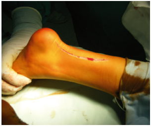
Fig 2. Slightly curved incision with medial concavity along the lateral aspect of the Achilles’s tendon relief

Fig. 3. The tendon is dissected and guided by locating the route of the future tenotomy
A. in equine club foot the Achilles’ tendon tenotomy is performed in a frontal “Z” incision.
B. in equinovarus club foot the Achilles’ tendon tenotomy is performed in a sagittal “Z” incision.
Fig. 4. The foot is guided to so as to form a 90 degrees position in relation to the ankle, suturing the tendon with the leg in slight hypercorrection

Fig. 5. Postoperatory, a cast holds the clubfoot still for 30 days, while it heals
Postoperatory, after the removal of the plaster cast, kinetotherapy is required in order to gain full recovery. Applying stress on the damaged foot, together with progressive walking, are allowed after 45 days.
Sometimes after surgery, in certain cases with pronounced spasticity, ankle braces are necessary. A close cooperaration with the neurologist is required, the specific treatment being controlled according to the future evolution of the patient (8).
MATERIAL AND METHOD
The study included 57 patients with the ages between 4 and 12 years, hospitalized at “The Paedia-tric Surgery Clinic” in Timisoara between 2000-2010 with spastic equine clubfoot, associated to child cerebral palsy.
After the surgery, these patients have been re-evaluated from the surgical point of view taking into consideration the two compulsory criteria of the normal locomotion: stability and motility.
DISCUSSION
Out of the 57 patients, 6 of them have shown absolute surgical contraindications, since they could not walk. A number of 8 patients have presented different degrees of psychic retardation, and in such cases cooperation pre and post surgery is difficult; that was why, the therapeutic results were modest.
We have to say that in this group, 7 operated patients have not been present to the subsequent controls, due to a weak cooperation with their families. In such cases by lack of a sound collaboration of all the entities involved and within medical team (the neurologist, physical therapist, surgeon), the initial good results, can be compromised.
The therapeutic evaluation was done according to a few criteria concerning the mobility and stability of the locomotion, which were clasified as:
- difficult walk – need support – 10 cases;
- a bit difficult walk – don’t need support – 5 cases;
- almost normal walk – 30 cases;
CONCLUSIONS
Therapeutic results in crooked leg in comorbidity with child brain paralysis depend on a good interdisciplinary collaboration of neurologist, physical therapist, and surgeon. The lack of such a collaboration can compromise the results achieved through surgery. Also, the evolution of the patient depends on a good cooperation surgeon-patient and surgeon – patient’s family.
BIBLIOGRAPHY:
- Bensahel H, Guillaume A, Czukonyi Z, DesgrippesY; Results of physical therapy for idiopathic club foot: A longterm follow-up study. JPediatr Orthop1990;10:189—92.
- DiméglioA; Classification et evaluation du pied bot varus équin. Monographie du GEOP: le pied de l’enfant. Édition Sauramps médical; Montpellier.2001,125—37
- Dobbs MB, Corley CL, Morcuende JA, Penseti IV; Late recurrence of club foot deformity: A 45 years follow up.Clin Orthop 2003, 411: 188- 92
- De Maio F,Orefice A, Ippolito E, Mancini F, Potenza V; Leg muscle atrophy in idiopathic congenital club foot. J Child Orthop 2008; 2 (Suppl.1): S38
- Herzenberg JE, Radler C, Bor N.; Ponseti versus traditional methods of casting for idiopathic club foot. J Pediatr Orthop 2002; 22:517—21
- Ippolito E, Fasetti P, Caterini R, Tudisco C ; Long-term comparative results in patients with congenital club foot treated with two different protocols. JBone Joint Surg 2003; 85 Am: 1286—94.
- Ponseti IV; Relapsing club foot: causes, prevention and treatment. Iowa Orthop J 2002; 22:55—6.
- Ponseti IV,Zhivkov M, Davis N, Sinclar M, Dobbs MB, Morcuende JA; Treatment of the complex idiopathic club foot. ClinOrthop 2006; 451:171—6.
- Niţu Monica, Locul kinetoterapiei în tratamentul piciorului varus equin, 2006
- Tachdijian’s Pediatric Orthopedics (the 3rd edition) Vol. 2.